


Endodontic retreatment is performed when a previously treated tooth requires further intervention due to persistent disease, recurrent infection, or other clinical findings. While retreatment can be technically demanding, careful case assessment, appropriate instrumentation, and effective cleaning protocols may help clinicians manage these challenges and improve the likelihood of achieving treatment objectives.
Every retreatment case presents unique anatomical and restorative considerations. Factors such as existing restorations, residual filling materials, calcified canals, and complex canal anatomy may influence clinical decision-making. A systematic approach allows clinicians to identify potential obstacles before treatment begins and select suitable techniques and instruments for each stage of the procedure.
Below are five commonly encountered challenges during endodontic retreatment and practical considerations for addressing them.
1. Skipping Coronal Assessment
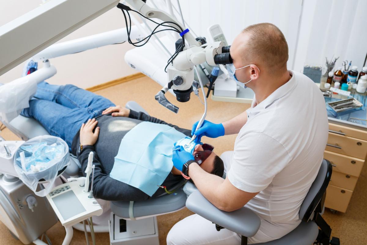
A successful retreatment begins before any instruments enter the canal system. Failing to evaluate the coronal portion of the tooth may result in overlooked restorations, cracks, fractures, or coronal leakage that could contribute to treatment complexity.
Careful clinical examination combined with appropriate radiographic assessment helps clinicians determine the restorability of the tooth, identify possible causes of previous treatment failure, and establish adequate access to the root canal system.
Clinical consideration:
- Assess existing restorations and coronal integrity.
- Evaluate for cracks, fractures, or recurrent caries.
- Create conservative but adequate access for improved visualization.
2. Incomplete Removal of Previous Filling Materials
Residual gutta-percha, sealers, carrier-based obturation materials, or other filling remnants may obscure canal anatomy and make negotiation of the original canal pathway more difficult.
Complete removal of previous filling materials may not always be possible in every clinical situation, but maximizing removal where clinically appropriate helps improve access for cleaning, shaping, and disinfection.
Clinical consideration:
- Select appropriate retreatment files and removal techniques.
- Use magnification when available.
- Reassess canal anatomy after filling removal before proceeding with instrumentation.
3. Missing Additional Canals
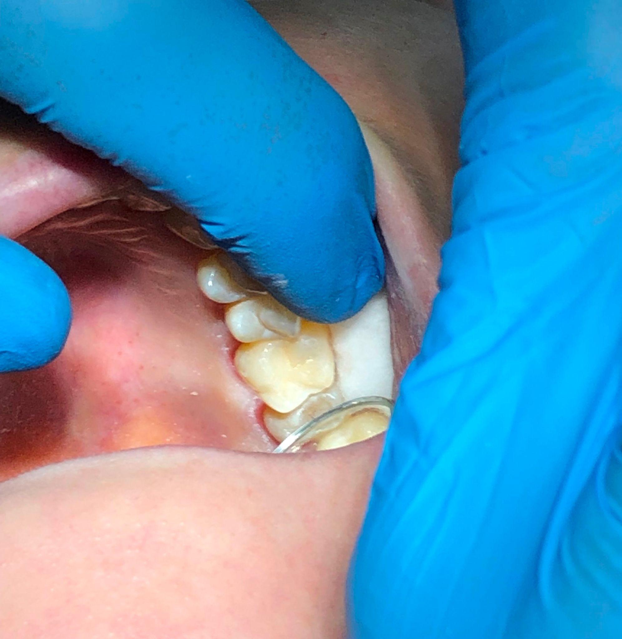
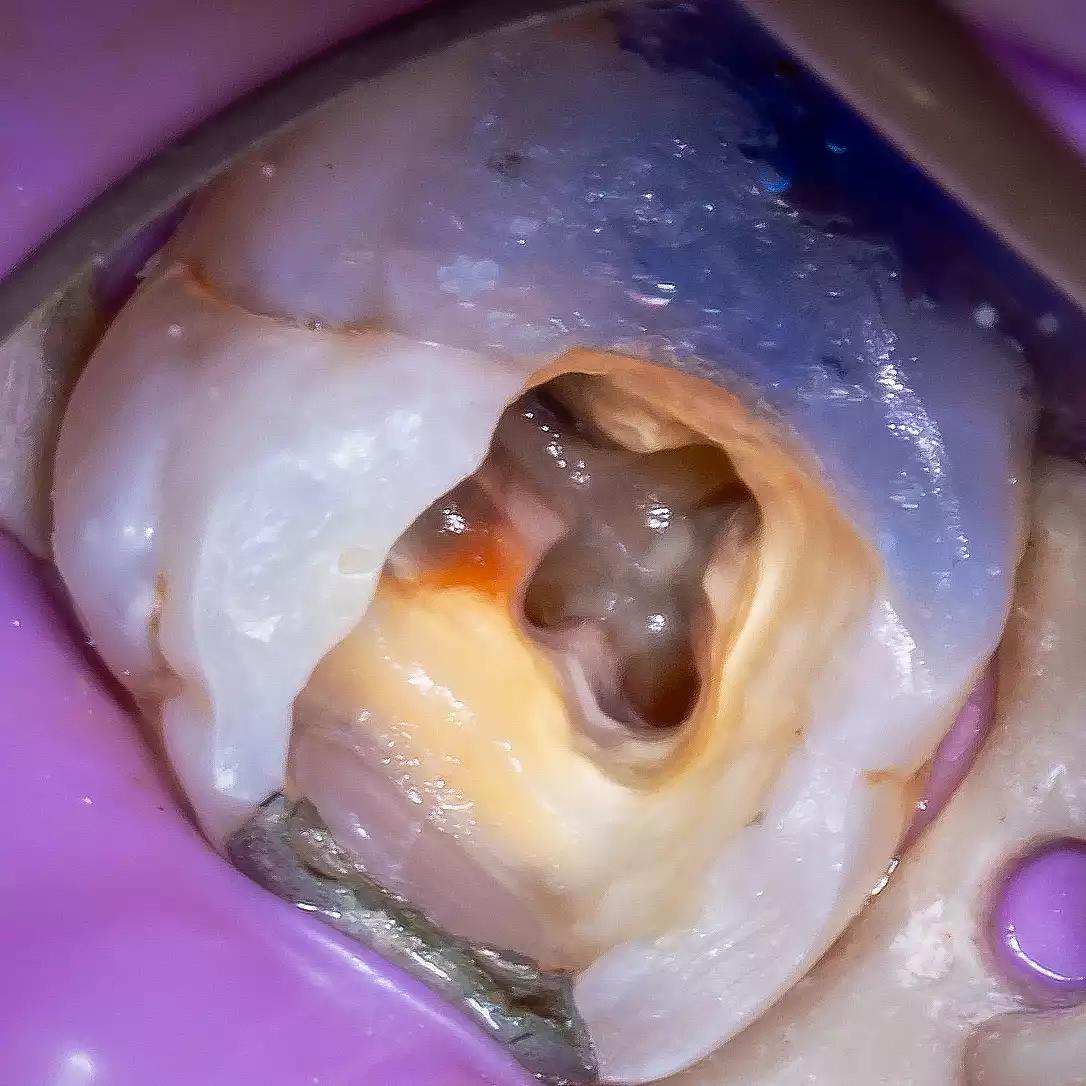
Complex root canal anatomy remains one of the most common challenges in endodontics. Untreated canals or anatomical variations, such as MB2 canals in maxillary molars or middle mesial canals in mandibular molars, may contribute to persistent disease if they are not identified during treatment.
Careful exploration of the pulp chamber floor, knowledge of root canal anatomy, and the use of magnification can assist clinicians in locating additional canals when present.
Clinical consideration:
- Review preoperative radiographs or CBCT imaging when clinically indicated.
- Inspect developmental grooves and anatomical landmarks.
- Use illumination and magnification to improve visualization.
4. Forcing Instruments Through Blocked or Calcified Canals
Negotiating calcified or obstructed canals requires patience and controlled instrumentation. Applying excessive force may increase the risk of procedural complications such as ledging, canal transportation, perforation, or instrument separation.
Establishing canal patency and developing a glide path before introducing larger shaping instruments helps support safer and more controlled instrumentation.
Clinical consideration:
- Begin with small hand files.
- Progress gradually while maintaining working length.
- Avoid excessive apical pressure during canal negotiation.
5. Underestimating Irrigation
Mechanical instrumentation shapes the canal system, but it does not reach every surface within the complex root canal anatomy. Irrigation plays an important role in flushing debris, dissolving organic tissue where appropriate, and reducing the microbial load within the canal system.
Proper irrigation protocols, combined with suitable activation methods when indicated, may enhance irrigant distribution throughout the canal system.
Clinical consideration:
- Use appropriate irrigants according to established clinical protocols.
- Ensure adequate irrigant volume and replenishment.
- Consider activation techniques where clinically appropriate.
Supporting Retreatment with Appropriate Endodontic Instruments
Endodontic retreatment often requires a combination of careful planning, sound clinical technique, and reliable instruments designed for different stages of the procedure. Depending on the clinical situation, clinicians may use dedicated retreatment files, hand files for canal negotiation, glide path instruments, rotary shaping systems, irrigation devices, and magnification aids to facilitate access, cleaning, and shaping.
Instrument selection should always be based on the clinician’s assessment of the individual case, the anatomical complexity of the tooth, and the intended treatment approach. Following the manufacturer’s instructions for use and established clinical guidelines helps support safe and effective instrument handling.
Conclusion
Endodontic retreatment is a meticulous procedure that requires careful diagnosis, thoughtful planning, and attention to detail throughout every stage of treatment. By recognizing common procedural challenges—including incomplete assessment, residual filling materials, missed canals, difficult canal negotiation, and insufficient irrigation—clinicians can adopt techniques that support more predictable treatment workflows.
While no single technique or instrument guarantees clinical success, combining sound endodontic principles with appropriate products and careful execution helps clinicians manage retreatment cases more effectively and efficiently.
